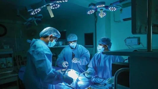
In a remarkable display of surgical expertise, doctors at a Mumbai hospital have successfully performed a complex, high-risk procedure on a 32-year-old woman diagnosed with a rare giant right sided diaphragmatic hernia.
The condition had led to the displacement of multiple abdominal organs into the chest cavity, resulting in critical compression of the right lung and a severe impact on her ability to breathe.
The woman had been experiencing persistent breathlessness, which had progressively worsened to the point where even minimal movement became difficult. Her medical history revealed a traumatic injury at the age of 12, considered a likely contributing factor.
Clinical evaluation, followed by advanced imaging, confirmed extensive herniation of the liver, stomach, duodenum and colon into the right side of the chest. This had caused a marked mediastinal shift along with significant compression of the right lung. Her morbid obesity further increased the complexity of the case.
Diaphragmatic hernias of this scale are extremely uncommon in adults and carry a high degree of surgical risk. In this instance, the diaphragmatic defect measured approximately 20 centimetres at its largest dimension. This allowed even the entire liver to migrate into the thoracic cavity, severely compromising respiratory function. There are very few cases, that is 173 documented in the world till date. In India this is the 1st operated case of Massive and giant diaphragmatic hernia of its kind in an adults with a defect of 20 cms.
Given the extent of organ displacement and the prolonged duration of the condition, the case required meticulous planning and close coordination across specialties. A multidisciplinary team led by Dr Vimesh Rajput, Consultant Thoracic Surgery and Dr Nilesh Doctor, Director (Administration) Surgical Gastroenterology undertook the surgery. Anaesthesia was managed by Dr Mohit, Consultant Anaesthesiology intensive care was led by Dr Shruti Tandon, Addl. Director Critical Care and physiotherapy support was provided by Dr Usha Kasare, Consultant Physiotherapy.
The procedure lasted nearly 10 hours. It was initially attempted laparoscopically but had to be converted to an open approach due to dense adhesions between the liver, diaphragm and lung. Through an abdominal approach, the surgical team carefully mobilized and repositioned the herniated organs back into the abdominal cavity. The diaphragmatic defect was kept open to be repaired via Thorax, while tight closure of the abdominal muscles was deliberately avoided to control intra-abdominal pressure and reduce the risk of abdominal compartment syndrome.
This was followed by a thoracic phase using video assisted thoracoscopic surgery, where the diaphragmatic hernia was repaired in two layers of Mesh to ensure structural stability.
Speaking about the case, Dr. Vimesh Rajput Consultant Thoracic Surgery at Jaslok Hospital and Research Centre said, “As the thoracic surgeon, my focus was on carefully dissecting the dense adhesions and repairing the large diaphragmatic defect. The organs had remained displaced for so long that restoring normal anatomy required extreme precision and patience. Using video-assisted thoracoscopic techniques, we ensured structural stability with 2 layered mesh placement. It was a demanding process, but seeing the patient regain her breathing capacity made every hour of the surgery worthwhile.”
Reflecting on the case, Dr. Nilesh Doctor Director- Administration of Surgical Gastroenterology explained, “My role was to mobilise and reposition the liver, stomach, and bowel, which had migrated into the chest cavity. The 20 cm diaphragmatic defect was massive, and repairing it required meticulous planning. We reduced the abdominal herniated contents back to abdomen, didn’t close the muscles of abdomen as it was difficult to close, instead we used mesh in the abdominal wall as well so that the intra-abdominal pressure must not rise. It was a complex challenge, but restoring the patient’s anatomy and laying the foundation for her recovery was deeply rewarding.”
The postoperative course remained critical and required prolonged intensive care support. The patient was on mechanical ventilation for 9 days and subsequently underwent a tracheostomy. Her recovery involved the management of pneumothorax and Respiratory care, which required advanced antibiotic therapy. Gradual respiratory rehabilitation along with nutritional support played a crucial role in stabilizing her condition and supporting recovery.
Despite the complexity of the surgery and the demanding postoperative phase, the patient showed steady improvement. Over nearly three weeks, she gradually resumed oral intake and regained mobility. At the time of discharge after 22 days of hospitalization, she was able to walk on a treadmill for 20 to 30 minutes without oxygen support.
Recounting her experience, the woman said, “I remember reaching a point where even breathing felt like an effort, and that fear stayed with me as I went into surgery. Waking up on support and then facing complications like infection was overwhelming, but the well-trained doctors and nurses were the reason for my healthier recovery. Walking on treadmill was an achievement for me where a normal breathing once was impossible.”
Mumbai doctors save life of woman unable to breathe due to massive 20 cm hernia